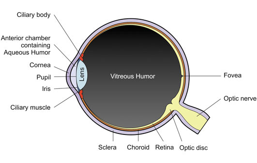
This is the white of the eye – the tough opaque outer coating. Six tiny muscles are connected to it, which control the eyes movement.
The Cornea is the transparent coating which covers the Iris and the Pupil at the front of the eye. The Cornea together with the Lens, helps refract the light and allows the eye to focus.
The Iris is the coloured part of the eye. Tiny muscles inside the Iris control the amount of light which can enter the eye by opening and closing the pupil.
Light enters the eye through the pupil. The size of the pupil is controlled by the Iris. When it is very bright and there is lots of light, the pupil is small. When it is dark, the pupil grows bigger to allow more light into the eye.
The purpose of the Lens is to focus light onto the retina at the back of the eye. Until we reach our mid 40s, the lens is able to alter shape, enabling the eye to focus on nearer objects.
The Retina contains millions of photoreceptors (sensors which convert light into electric impulses which are sent along the optic nerve to the brain). There are two types of photoreceptors – Rods (which allow us to see in low light) and Cones (which allow us to see colour and work best in brighter light).
The Optic Nerve send signals representing colour from the eye to the brain, which then interprets what we see. Where the Optic Nerve leaves the retina there are no sensory receptor cells, meaning we have a blind spot in our eye.
When we blink, the tears lubricate, nourish and protect the front of the eye.
Tears have three main components: The lachrymal gland produces a watery component glands in our eyelids produce an oily component, while other cells produce a mucus. These mix together to create a film which covers the white of the eye and the cornea. When we blink, the film is wiped across the eye by the eyelids.
If insufficient tears are produced or the constituents are out of balance it can result in sore, dry eyes.
Wearing glasses or sunglasses can help to reduce tear evaporation.
Instilling artificial tears can also help. In some cases, the tears can be kept in the eye for longer by plugging the tear ducts.
In a normal eye, light is focussed by the cornea and the lens to form a sharp image on the retina at the back of the eye. As we get older, the lens can become hazy.
This is known as a cataract. This makes the vision rather blurred or hazy even when wearing the correct spectacles.
In most cases, vision can be restored by a simple operation.
The eye is numbed and a special tool is used. This tool removes the cloudy part of the lens. A plastic lens designed for your eye is made. This is rolled up so that it can be inserted into the eye and placed in the lens capsule, where it springs into position. Usually, the new clear window greatly improves the quality of vision.
In a normal eye, light entering the eye is focussed by the cornea and the lens to form a sharp image at the back of the eye on the retina. Astigmatism occurs when the cornea or lens inside the eye is slightly “rugby ball shaped”, causing a distortion of focus. The effect of this is that lines of different orientations come to a focus at different points in the eye. This results in blurred vision when looking in the distance and reading. The degree and angle of astigmatism can vary from eye to eye.
If you have astigmatism you may notice that objects of certain orientations look clearer.
The eye is filled with a jelly-like substance known as the vitreous. This jelly is loosely attached to the retina at the back of the eye. As we get older, the jelly tends to shrink a little, and can tug on the retina. In some cases the vitreous can detach from the retina. As it tugs on the retina, this can cause flashing lights in the corner of your eye followed by the appearance of floaters which move across your vision.
In a small percentage of cases, a vitreous detachment can result in a retinal detachment, which is potentially serious. Therefore, if you are experiencing these symptoms, you should seek immediate attention from an Optician or GP and not drive to your appointment.
The retina is quite fragile and can tear. This may happen spontaneously or following a blow to the eye or head. As the retina tears, flashing lights are often seen – usually out of the corner of the eye. If this is not treated, fluid can pass through the tear and cause a larger area to become detached. This can lead to a permanent loss of vision if not treated straightaway. A tear or a detachment often causes a sudden increase in the number of floaters.
If you are experiencing these symptoms, you should seek urgent attention from an Optician or GP and not drive to your appointment
When light hits the back of the eye, tiny cells convert the light into nervous impulses.
These travel across the retina, through the optic disc, to the brain. Glaucoma is a group of diseases which cause these nerves to become damaged. When this happens the nerves become unable to send the impulses which leads to blind spots, initially out of the corner of the eye. If the condition is not treated, the blind spots get larger and can lead to blindness. It is known that pressure in the eye is an important factor in this condition. The production and drainage of “water” in the front of the eye affects the pressure. If this gets out of balance the pressure in the eye can increase. Glaucoma can be controlled by using eye drops and/or laser treatment to reduce the pressure in the eye.
Light is focussed on the retina at the back of the eye. The macula is a small area in the centre of the retina that we use to read and see fine detail. The macula consists of several layers of tiny cells. As we get older, these cells can fail to function properly leading to a build up of deposits and a loss of cells.
This can lead to distortion and over time, it can become difficult to read and see fine detail. The condition usually progresses quite slowly and various aids are available to help you to see smaller print.
Light entering the eye is imaged on the retina at the back of the eye. The macula is a small area in the centre of the retina. This is the part of the eye that we use to read and see fine detail. The macula consists of several layers of tiny cells.
As we get older, some of these can fail to function properly. This can lead to a build up of deposits in the retina and the growth of new blood vessels. These new blood vessels are fragile and bleed easily. If this happens, there may be a sudden loss of central vision and objects may appear distorted.
If you are experiencing these symptoms, you should seek urgent attention from an Optician or GP and not drive to your appointment.
Diabetes can have various affects on the eyes, most commonly the retina. The walls of the blood vessels tend to become weakened. Over time, the walls may bulge – known as an aneurysm. This creates an ‘eddy’ in the blood flow which can eventually block.
Blood and other fluids also tend to leak from the blood vessels. This results in a build up of lipids in the layers of the retina. Because of the reduction in blood flow, new blood vessels tend to form. However these blood vessels are fragile and bleed easily. If the new blood vessels bleed into the jelly in the eye (the vitreous) there can be a sudden loss of vision. But this may clear somewhat over time.
or if you have any questions please contact us by filling the from below or give us a call on 01379 650899