We are proud to be the first practice in the area to offer both 3D-OCT imaging and Digital Retinal Photography.
We are proud to be the first practice in the area to offer both 3D-OCT imaging and Digital Retinal Photography. Both of these techniques are particularly relevant to anyone with risk factors such as family history of eye problems or sudden visual changes. However, anyone can benefit from the extra peace of mind of having the most comprehensive eye care that technology can provide, in addition to our comprehensive eye examination.
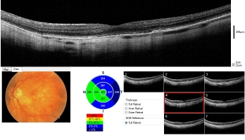
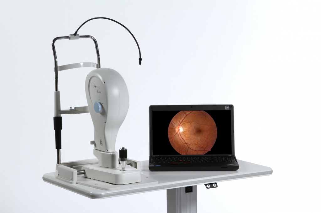
OCT (Optical Coherence Tomography) is the latest technique for the imaging of the internal structures of the eye. The technology is best likened to a 3D MRI scanner for the eyes. This allows us to actually see below the surface of the retina and view the microscopic layers beneath. It is currently commonly used in Hospital Eye Departments for the diagnosis and management of many common eye conditions. However, only a minority of Optometric practices are able to offer the benefits of OCT to their patients.
The process is very quick and totally painless, without any bright flashes. We can normally obtain all the measurements we require without the need for dilating drops. Therefore, this service is ideal for those driving to their appointment.
By viewing the different layers of the retina, we can detect and differentiate between wet and dry age related macular degeneration (AMD) and numerous other retinal conditions, including diabetic maculopathy. This allows us to determine whether urgent hospital attention is required or when just monitoring is required.
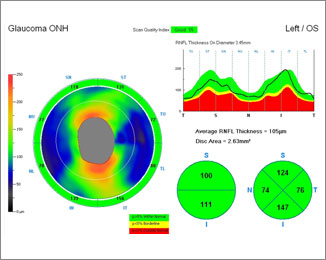
The OCT can also measure the retinal thickness in key areas of the eye, which are susceptible to damage from glaucoma. By comparing these findings to databases of known normal individuals and by detecting subtle changes to these areas over time, OCT can give us an early warning of glaucoma. The aim is to detect potential glaucoma before any damage to vision. Conventional eye examinations normally detect this condition at a later stage, by which time irreversible visual loss may have already occurred
.
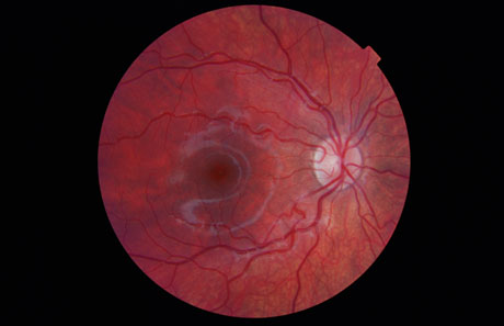
Put simply, digital retinal photography allows us to capture a snapshot of the appearance of a large portion of retina in one go. This in turn, means that we can look at the ‘big picture’ of the back of the eye, rather than visualising small portions at any one time. By storing and comparing these images, we can detect any subtle changes over time, which may otherwise be missed by more conventional means. The quality of the images obtained is dependent on pupil size. This means that we often have to dilate your pupils using drops, which can temporarily make your vision blurry. Please bear this in mind if you are planning to drive to your appointment and allow plenty of extra time and someone else to drive if possible.
or if you have any questions please contact us by filling the from below or give us a call on 01379 650899